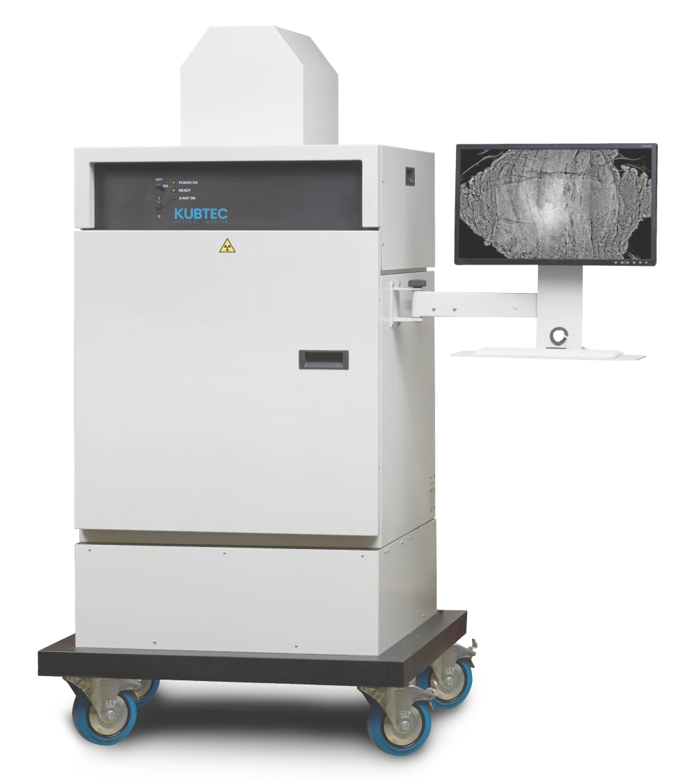
XPERT 80-L

Razor-sharp X-ray images for pathology

Working quickly and accurately in the cutting room starts with good imagery. With the XPERT 80-L Specimen Radiography System of KUBTEC pathologists and pathology assistants gain immediate insight into the tissue they are examining. Thanks to razor-sharp X-ray images as well as optical images, this system shortens turnaround time and increases precision in every section.

Largest detector in the pathology market

KUBTEC is momentarily the only provider in pathology that delivers a detector size of 17” x 17” (43 x 43 cm) . This provides ample space for large specimens such as organs, bones or a full-term foetus. This makes the XPERT 80-L system unique in its class.

Want to experience the XPERT 80-L for yourself? Fill in the contact form below.

Learn more

    Send

    Key features

    • 17” x 17” (43 x 43 cm) detector: unique in pathology, suitable for large specimens

    • HD optical camera: for accurate visual orientation of the specimen in real time

    • Automatic Specimen Alert: alerts when a specimen is left in the device

    • Image Blender™: combines optical and X-ray images for a complete anatomical view

    • Density Profile: graphical representation of differences between calcified and non-calcified bone

    • Copy Path: easy export of images to the Laboratory Information System (LIS)

    One image, all information

    Thanks to the “Image Blender”, optical and X-ray images can be seamlessly combined. This allows you to locate at a glance:

    • Surgical clips

    • Resection margins

    • Microcalcifications

    You simply determine (before cutting) which tissue parts should be placed in the cassettes. With the XPERT 80-L system, you see more, faster and more precisely. You make targeted choices, save time and increase the quality of the diagnostic process.

    linkedin facebook pinterest youtube rss twitter instagram facebook-blank rss-blank linkedin-blank pinterest youtube twitter instagram